

More than 80% of plants on Earth partner with mycorrhizal fungi to extract nutrients from soil.
Underground, in a zone of soil known as the rhizosphere, these fungi grow into vast networks of tubular cells called mycelium (individual cells are called hyphae). These tiny hyphae—about one tenth to one hundredth the width of a human hair—can gather nutrients in the soil and send them into plants.
A network of hyphae is seen under a microscope, magnified 630 times.
If plants don’t get enough nutrients, neither do the animals that depend on them or the animals that depend on those animals. Furthermore, some research has suggested that mycorrhizal fungi absorb the equivalent of 13 billion tons of carbon dioxide annually.
All of this makes the rhizosphere essential to the health of our planet and its inhabitants.
“Basically everyone on Earth is dependent on this really, really tiny zone around roots, in a way.”
“Basically everyone on Earth is dependent on this really, really tiny zone around roots, in a way,” said Henri Braunmiller, a soil ecologist and graduate student at the Technical University of Munich.
Again, hyphae are tiny: One gram of soil can contain up to 90 meters of mycelium. Most of the time, imaging them requires removing them from soil, which means scientists don’t have a 3D view of what the interconnected networks of roots, hyphae, and fungal spores look like.
But in new research published in New Phytologist, Braunmiller and his colleagues report a way to study this network in its natural environment for the first time using a technique called synchrotron computed tomography (CT).
Justin Stewart, an ecologist with the Society for the Protection of Underground Networks and Vrije Universiteit Amsterdam, told Eos in an email that the capacity to study fungal hyphae within intact soils at higher resolution represents a “major advance.”
“This makes it possible to examine the interface between living fungal networks and the mineral soil matrix, and opens new avenues for studying how fungal-derived carbon is transferred to soil minerals,” he said.
Shedding Some Light
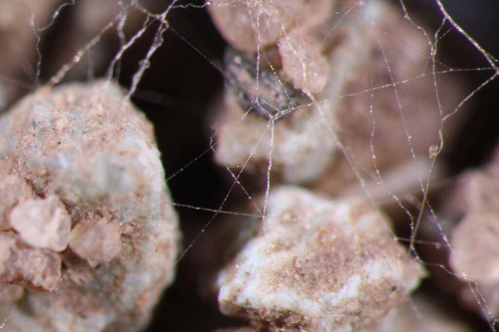
Hyphae are seen stretching between soil particles in a petri dish. This light microscopy image, taken by paper coauthor Jan Jansa, measures less than 1 millimeter across.
Researchers can learn about mycorrhizal networks when they form in a petri dish like in the above photo or in soil chips, which are silicon wafers designed to mimic the structure of soil. But the information scientists gain from these methods is limited because the rhizosphere’s many facets are not replicated in these environments.
That’s where CT, the same technology used in medical scans, comes in. A typical laboratory CT, especially a high-resolution model, can take hours to image a sample, however. In those hours, fungi move and grow, blurring the image.
“A synchrotron produces a million times the energy that your lab CT produces.”
So the team turned to a synchrotron, a specialized particle accelerator that can create highly intense beams of light.
“A synchrotron produces a million times the energy that your lab CT produces,” Braunmiller said. “So you don’t want to stand in the way of the beam. But then you can do the same images in a couple of minutes.”
Braunmiller and his team set out to grow tomato plants in both loam and sandy soils containing fungi spores, hyphae, and colonized root fragments.
Then, they added small cylinders of soil, about 12 millimeters across and 60 millimeters tall, to the pots. The cylinders acted as baskets, covered in holes big enough—2 by 4 millimeters—for both the hyphae and plant roots to grow into. When removed after about 8 weeks of tomato growth, the cylinders contained a cross section of the rhizosphere’s various elements.
In this microscopy image, fungal structures, stained blue, are growing inside the root of a tomato plant.
The team drove the samples 9 hours from Munich to SOLEIL just outside of Paris, one of France’s very large scale research infrastructures and home to one of only a few dozen existing synchrotrons.
It was after midnight by the time researchers began to scan the samples. When the team looked through the images in succession, they saw tiny white points moving across the image.
They were hyphae.
“It was so exciting, because nobody was expecting that we could see them, because they’re so tiny,” Braunmiller said. “They’re right at resolution edge.”
Understanding a Tool
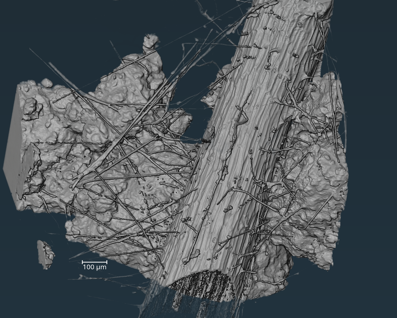
By stacking the images they’d gathered using CT, Braunmiller and his team could reconstruct 3D images of the full rhizosphere in its natural environment for the first time.
A 2D image (right) can be seen as the cross section of a 3D depiction (left).
In the left image, the large cylinder is a root, the thin filaments are hyphae, and the surrounding blobs are soil aggregates.
The researchers expanded their visualizations from there.
For instance, adding colors allowed them to conduct image analysis on the volume and surface area of the hyphal network.
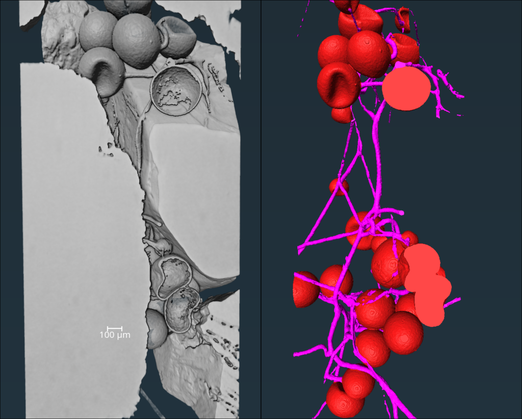
Here a 3D reconstruction shows hyphae (pink) and fungal spores (red) growing in sandy soil.
Visualizing these networks is important because of the crucial role the rhizosphere plays in plant health, food systems, and the carbon cycle.
The team hopes their findings could be particularly useful for helping plants get enough water during drought, especially as drought increases in the face of climate change. Mycorrhizal fungi can help plants with nutrient uptake and pathogen resistance, so using these networks strategically could help improve crop yield stability and reduce dependence on herbicides and fungicides.
These networks “are a tool, but you also have to understand the tool,” Braunmiller said, explaining that these networks are more helpful in some settings than others, and researchers don’t fully understand why. “We’ll look also at how these networks work in different kinds of soils, so we can give recommendations on when to use them and when to maybe not.”
—Emily Gardner (@emfurd.bsky.social), Deputy Editor
Correction: This article was updated on 1 October to correctly describe soil aggregates in a photograph.